
What is Gestational Trophoblastic Disease ?
Gestational Trophoblastic Disease (GTD) refers to uncommon medical conditions that occur due to abnormal proliferation of trophoblastic tissue that normally forms the placenta during pregnancy. The cells in this system fail to produce a functioning placenta, which would enable fetal development; instead, they continue to multiply unchecked in the uterus. Some GTD variants show harmless behavior, which requires minimal treatment, whereas other variants demonstrate cancer-like behavior, which needs whole-body chemotherapy treatment.
GTD stands out from other cancers because it links directly to pregnancy while serving as a highly treatable cancer when doctors use proper testing and treatment methods. Chemotherapy proves effective even for patients whose cancer cells spread throughout their body to remote organs. Doctors can achieve high survival rates through proper patient monitoring and established medical check-up routines.
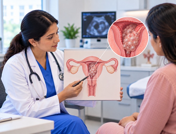
GTD can occur after any pregnancy event — including a full-term pregnancy, miscarriage, ectopic pregnancy, abortion, or molar pregnancy. The process of diagnosis and effective treatment requires early identification of unusual symptoms together with ongoing observation of β-hCG levels.
What is the difference between a Hydatidiform Mole and Gestational Trophoblastic Neoplasia?
Gestational Trophoblastic Disease encompasses various medical conditions, including non-cancerous tumors and cancerous growths. Medical specialists identify these health conditions by using two main medical categories, which include the following:
Hydatidiform Mole (Molar Pregnancy)
The most prevalent form of this condition is premalignant. The condition further divides into two main groups, which include:
- Complete mole
- Partial mole
When a sperm fertilizes an empty egg, aberrant placental tissue grows without fetal development, resulting in a full mole. This occurs when two sperm fertilize a normal egg, resulting in abnormal fetal and placental tissue.
Some cases of molar pregnancy develop into invasive disease despite successful treatment through uterine evacuation in most cases.
Gestational Trophoblastic Neoplasia (GTN)
This group includes malignant forms:
- Invasive mole
- Choriocarcinoma
- Placental site trophoblastic tumor (PSTT)
- Epithelioid trophoblastic tumor (ETT)
Choriocarcinoma represents the most dangerous cancer type, which shows a strong reaction to chemotherapy. The rare tumors called placental site tumors and epithelioid trophoblastic tumors need doctors to perform surgical treatments.
What are the Symptoms & Causes of Gestational Trophoblastic Disease ?
Causes of Gestational Trophoblastic Disease
Genetic abnormalities in trophoblast cells result from aberrant fertilization, which causes GTD. A partial molar pregnancy happens when there is an excess of the father's chromosome material, which results in aberrant tissue development. A complete molar pregnancy happens when sperm fertilizes an empty ovum, producing an ovum that has all of the father's chromosomes.
The following factors increase risk:
- Maternal age below 20 or above 35
- Previous molar pregnancy
- History of miscarriage
- Nutritional deficiencies (rarely implicated)
Most people develop the disease without known reasons despite these established connections.
Symptoms of Gestational Trophoblastic Disease
Manifestations usually occur in early stages of pregnancy and can be confused with miscarriages.
These are some common manifestations:
- Vaginal bleeding in the first trimester
- Excessive nausea and vomiting (Hyperemesis gravidarum)
- The uterus is growing faster than the gestational age.
- Lower pelvic pain
- Discharge of cysts or grapelike substances
Advanced cases show symptoms which include:
- Persistent bleeding after pregnancy
- Elevated or rising β-hCG levels
- Shortness of breath (lung metastasis)
- Severe headache or neurological symptoms (brain involvement)
- Abdominal pain due to liver metastasis
The symptoms of GTD cause the body to produce excessive β-hCG, which creates strong hormone-related symptoms.
How is Gestational Trophoblastic Disease Diagnosed & Tested ?
Early and accurate diagnosis is essential.
Beta-hCG Testing
The major tumor marker for GTD is represented as β-hCG. The values of this tumor marker are considerably higher than those found in a normal pregnancy. The presence of increasing hCG levels after uterine evacuation suggests the development of malignant cells.
Ultrasound Examination
The pelvic ultrasound in GTD usually demonstrates:
- “Snowstorm” or Cluster of Grapes appearance in a complete mole
- Abnormal Fetus with cystic Placenta in a partial mole
- Absence of a viable embryo in many cases
The ultrasound examination helps doctors establish a treatment plan before surgeons proceed with the surgical operation.
Histopathological Confirmation
Physicians use a microscope to examine tissue samples to establish the medical diagnosis after the patient undergoes a uterine evacuation.
Imaging in the Case of Cancer
In case of persistent GTN:
- CXR/CT scan (common metastatic organ - lungs)
- Abdomen and pelvis CT scan
- Brain MRI (in case of neurological symptoms)
These investigations will help in staging the disease.
What are the Stages & Risk Assessment for Gestational Trophoblastic Disease ?
The medical experts stage GTD based on the distance the disease has spread through the body:
- Stage I: Confined to the uterus
- Stage II: Spread to the pelvis
- Stage III: Lung involvement
- Stage IV: Distant metastasis (brain, liver, others)
The prognostic scoring system uses these factors for assessment:
- Patients age
- Previous pregnancy type
- Time elapsed since last pregnancy
- Current β-hCG measurement
- Size of tumors
- Locations of cancer spread
The system classifies patients into two groups:
- Patients with a low risk
- Patients with a high risk
The system determines the chemotherapy regimen required for each patient.
How is Gestational Trophoblastic Disease Treated ?
It requires implementing treatment plans tailored to the disease category and the patient's risk level.
The main treatment for Hydatidiform Mole involves:
D&C (suction evacuation and curettage) serves as the primary treatment method.
The process removes abnormal tissue through D&C from the uterus after the procedure is finished.
The medical staff performs weekly monitoring of β-hCG levels until the patient reaches normal values.
The medical team recommends birth control methods to patients from the time of treatment until their beta-hCG testing schedule for impending tests.
The majority of patients will achieve complete recovery without needing additional medical procedures.
Treatment of Low-Risk GTN
GTN patients with low risk receive treatment through methods that require monitoring of their β-hCG levels.
The medical team will administer single-agent chemotherapy, including methotrexate and actinomycin-D, if the patient shows persistent or rising β-hCG levels.
The two medications provide effective treatment results.
In cases of low-risk disease, the treatment achieves nearly 100 percent success.
The Treatment of High-Risk GTN
High-risk or metastatic disease requires:
- The application of multi-agent chemotherapy protocols
- Continuous patient observation at specialized oncology medical facilities
The treatment succeeds for patients with lung, brain, or liver metastasis since their tumors show an exceptional response to chemotherapy treatment.
Surgical Management
The surgical team considers a hysterectomy as an option for women who:
- Have finished their childbearing years
- Have a localized disease that persists.
- Experience persistent severe bleeding
The two types of tumors, which originate from placental sites and epithelioid trophoblast cells, show low response to chemotherapy treatment; therefore, surgical intervention is needed for their management.
Radiation Therapy
Radiation therapy is needed in rare situations to treat:
- Metastatic tumors in the brain
- Localized tumors that do not respond to chemotherapy
What are the side effects of GTD treatment?
The treatment produces these side effects:
The temporary Side Effects:
It includes:
- Nausea and vomiting
- Fatigue
- Hair thinning
- Mouth ulcers
- Low blood counts
- Increased risk of infection
The permanent side effects include:
- Rare fertility impairment
- Early menopause (uncommon)
- Rare secondary cancers
- Emotional distress
Modern treatment protocols prevent most patients from experiencing long-term medical problems.
Fertility & Future Pregnancy
The preservation of fertility is one of the most reassuring aspects that GTD treatment offers to patients.
Most women achieve three outcomes after treatment, which include:
- Resumption of their regular menstrual periods
- Natural conception after treatment
- Delivery of healthy babies
Patients should avoid pregnancy for 6 to 12 months during their monitoring period.
The prospects for future pregnancies are successful.
What is Outlook / Prognosis for Gestational Trophoblastic Disease ?
The medical field considers gestational trophoblastic disease among the cancers with the highest cure rates.
- Hydatidiform Mole: Virtually 100% survival rate
- Low-Risk Gestational Trophoblastic Neoplasia: Nearly 100% cure rate
- High-Risk Metastatic Gestational Trophoblastic Neoplasia: Over 85-90% cure rate
There are three factors that determine the outcome: early cancer diagnosis, adherence to follow-up, and prompt treatment.
Why is structured long-term follow-up necessary after Treatment?
Follow-up needs to be structured because it plays a critical role in healthcare operations.
The process includes:
- Weekly β-hCG testing until normal
- Monthly monitoring for at least 6-12 months
- Reliable contraception during surveillance
- Imagine if tumor marker levels rise.
Most recurrences occur within the first year and are treatable.
Emotional & Psychosocial Considerations
Patient's experience:
- Bereavement caused by miscarriage
- Stress caused by a possible cancer diagnosis
- Worry about future conception.
- Mental distress from continuous monitoring
In the care process, counseling and family involvement are necessary.
Why choose Tender Palm Super-Speciality Hospital for Gestational Trophoblastic Disease Treatment in Lucknow, India?
Tender Palm Super-Speciality Hospital offers advanced Gestational Trophoblastic Disease treatment in Lucknow, India, at an affordable cost. We have a team of experienced gynecological oncologists and surgical specialists who provide accurate diagnosis and both non-surgical and surgical treatment options including chemotherapy, uterine evacuation, and comprehensive hormonal management procedures. Our Gynecological Oncology and Surgical Care team has decades of experience in successfully treating Gestational Trophoblastic Disease in Lucknow, India.
To seek an Expert consultation for Gestational Trophoblastic Disease Treatment in Lucknow, India:
Call us at +91-9076972161
Email at care@tenderpalm.com
Our Experts
Awards & Accreditations


